Challenging our conventional management with the aid of 3D+
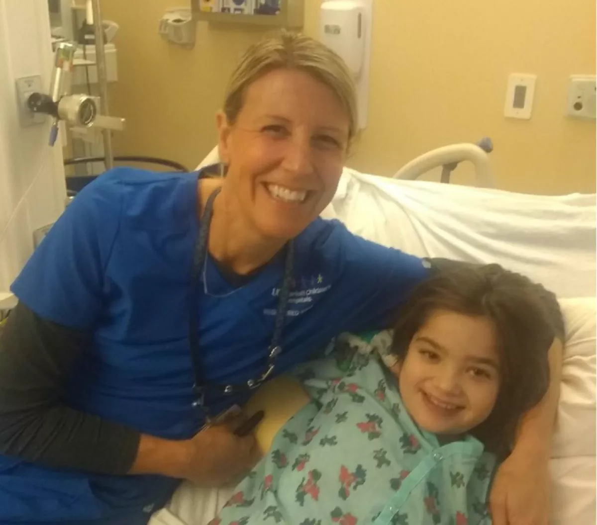
One of our patients is a 12-year-old girl with heterotaxy syndrome and complex congenital heart disease with complete atrioventricular septal defect. Her care was managed via the single ventricle pathway, which consists of a series of surgeries culminating with the Fontan procedure. This results in a functional single ventricle (pumping chamber) that pumps blood to the systemic circulation, while the pulmonary circulation is completely separate from the heart. Single ventricle palliation is a life-saving surgery for many forms of congenital heart disease. However, the long-term outcomes are not favorable, with significant morbidity and mortality in adulthood. Unfavorable long-term outcomes include: heart failure requiring heart transplants; arrhythmias requiring pacemakers/defibrillators; or other multisystem organ dysfunction.
Our patient was managed with this surgical strategy because it was previously felt that her heart disease was too complex to risk a two-ventricle repair. However, with the availability of 3D+ technologies, we re-investigated her case to determine the feasibility of a two-ventricle repair. We created digital and printed 3D models to aid the Pediatric Heart Center team and our surgeons with pre-procedural planning and surgical simulation. After re-investigating her anatomy with the 3D models, Dr. Reddy, chief of Pediatric CT Surgery, felt that he could successfully perform a two-ventricle repair. The patient underwent surgery in November 2019. Following her recovery, her parents reported that the patient had increased energy and was able to discontinue some of her cardiac medications. She continues to do well clinically.
Since the founding of CA3D+, we have re-evaluated several of our single-ventricle patients to determine if they, too, could undergo biventricular conversions. 3D+ technologies have been a game-changer for the management of these complex patients, offering them renewed hope of achieving better outcomes.
Following her recovery, the patient's parents reported that she had increased energy and was able to discontinue some of her cardiac medications. She continues to do well clinically.
When less is more
Our next patient was born with very complex heart disease consisting of an “upstairs downstairs” heart, double-outlet right ventricle, large ventricular septal defect (VSD) and pulmonary atresia. We created a 3D-printed model to plan a complex two-ventricle repair. However, during the process of modeling, we discovered a second VSD – a hole between the pumping chambers of the heart. The 3D model showed this VSD was in a location that would make it very challenging to close successfully, further complicating a risky neonatal surgery. Thus, we opted for a safer approach and performed a palliative procedure for her first surgery instead of a two-ventricle repair. To date, she has had two planned palliative surgeries and our plan is to perform a comprehensive two-ventricle repair when she is older. Over the years, we have implemented 3D+ technologies successfully to perform high-risk, complex, two-ventricle repairs, but in this case the 3D model pointed us toward a more conservative approach, which truly customized the surgical management to the patient’s unique anatomy. The young patient is now doing very well clinically.
Over the years, we have implemented 3D+ technologies successfully to perform high-risk, complex, two-ventricle repairs, but in this case the 3D model pointed us toward a more conservative approach.
“Redefining possible” in action
The first open fetal surgery in the world was performed at UCSF in 1981, and UCSF remains one of the leading centers for high-risk fetal interventions. Recently, the Pediatric Surgery and ENT Surgery teams called on CA3D+ to assist with an extremely high-risk fetal procedure. The patient was a fetus at 29 weeks gestation with a very large teratoma (tumor) arising from her chest and neck. There was significant concern that the tumor was compressing her airway, and the surgical teams were asked to evaluate the case for a potential EXIT procedure.
EXIT, or ex-utero intrapartum treatment, is a specialized surgical delivery procedure used to deliver babies who have airway compression. Prior to arriving at UCSF, the family had been evaluated at several other institutions, but no procedures had been offered due to the severity of the condition. Our surgical teams were deliberating about offering an EXIT procedure when Dr. Hanmin Lee, chief of Pediatric Surgery, consulted us at CA3D+ to assist with surgical planning. We obtained a fetal MRI and created a 3D model to show the anatomy of the fetus and the tumor. Specifically, we were able to identify a fetal airway (breathing tube) that the team could potentially access for a successful EXIT procedure. The surgical team informed us that, prior to 3D modeling, this crucial detail had not been clear. Given this information, we decided to proceed with an EXIT procedure, which was performed successfully in June 2021. The anatomy shown by the 3D model was verified to be accurate during the procedure, and the baby was delivered successfully. Subsequently, the teratoma was surgically resected, and the baby recovered well in our neonatal intensive care unit. Speaking with the surgical and intensive care team afterward, there was a consensus that the 3D model was extremely helpful in planning and executing this extremely high-risk, life-saving procedure. The young patient was recently discharged from the NICU, and to date she is doing well at home.
Preparing for the case
Dr. Shafkat Anwar, CA3D+ Medical director (2nd from left), and the EXIT team; Dr. Dylan Chan (left) and Dr. Hanmin Lee (center) prepare for the case.
Utilizing CA3D+ technology for more precise surgery, offering patients understanding and peace of mind
Another patient had a cancerous tumor in her right breast, and she was to undergo a partial right mastectomy. Michael Bunker, 3D+ engineer, made a 3D model of her entire breast, with the tumor visible inside. It was used both in surgical planning and during the procedure in the operating room to reference the location and size of the cancerous tissue to aid in ensuring that the tumor was completely removed. The patient was shown the model before her procedure and was delighted that she was able to see the tumor and could really understand what the surgeons were going to do. After her procedure, she remarked that being able to see and hold the model was extremely useful to understanding her own anatomy. She said, “I thought I had two separate tumors. The 3D model gave me clarity, showing the size of the one tumor and its true size.” She added, “Great work; we all need to know about the 3D technology. Visuals always make things clearer.”
Models for breast tumor patient
Dr. Tatiana Kelil, CA3D+ co-director (right), and Dr. Rita Mukhtar, breast and general surgeon
Allowing surgeons to visualize for reconstruction
This patient had a left ischium angiosarcoma (cancerous tumor of the blood vessels and lymph node vessels) develop on his pelvis. His orthopaedic surgeons determined it would be beneficial to utilize 3D modeling to better see the size, shape and location of the tumor, together with nearby bones and vasculature. Since the tumor hugged the unique shape of the hip bone, it was useful to show where the bone was underneath and inside the tumor.
By creating this model with unique color schemes, the surgical team was easily able to visualize the tissue, bone and vessels. In this case: arteries are red, vessels are blue and bones are white; the dark gray is a metal hip replacement; and the tumor itself is transparent opaque to yellow. Orthopaedic surgeon Rosanna Wustrack, MD, was able to perform a wide resection to remove the tumor and surrounding tissue. In her follow-up comments, she said, “This was a great model and allowed me to better understand the complex anatomy and position of the tumor.”
“This was a great model and allowed me to better understand the complex anatomy and position of the tumor.”


